Level 12 Gelenke, Knochenpunkte etc, Memrise
The internal acoustic canal (IAC) , also known as the internal auditory canal or meatus (IAM), is a bony canal within the petrous portion of the temporal bone that transmits nerves and vessels from within the posterior cranial fossa to the auditory and vestibular apparatus. Gross anatomy

EXTERNAL EAR part 2 External Acoustic Meatus YouTube
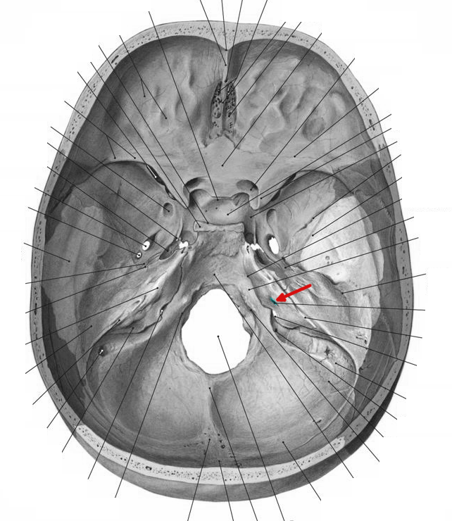
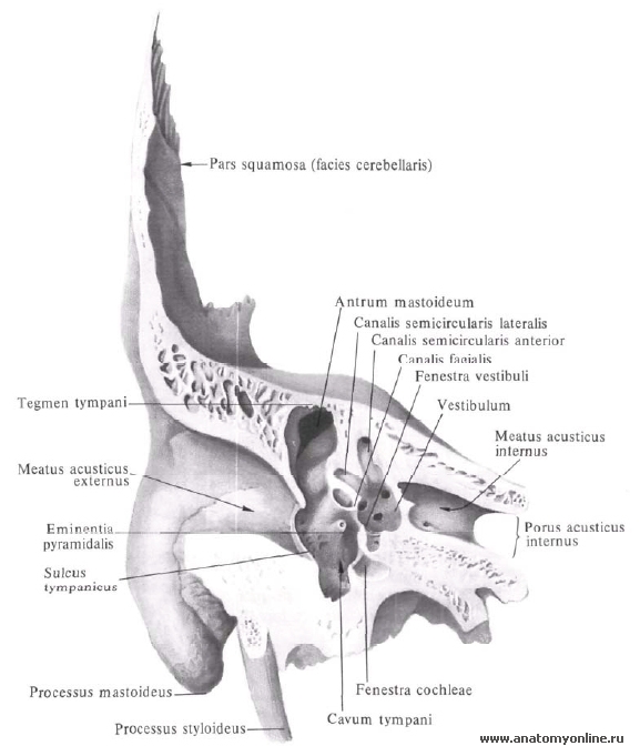
Definition The internal acoustic opening is a large orifice near the center of posterior surface of petrous part, its size varies considerably; its margins are smooth and rounded, and it leads into a short canal, the internal acoustic meatus, about 1 cm. in length, which runs lateralward.
External Auditory Meatus/Acoustic Meatus Earth's Lab
The influences of porus acusticus internus on ethnicity and importance in preoperative and intraoperative approaches. R. Şekerci Eren Ogut N. Keles-Celik. Medicine.. Amac: Bu calisma 18-60 yas arasi saglikli Turk populasyonunda meatus acusticus internus morfometrisini belirlemek amaclandi, yurutulen bu calisma retrospektif bir calismadir.

Meatus Anatomy
The internal auditory meatus (also meatus acusticus internus, internal acoustic meatus, internal auditory canal, or internal acoustic canal) is a canal within the petrous part of the temporal bone of the skull between the posterior cranial fossa and the inner ear . Structure
Divertikel Meatus acusticus internus pacs
Meatus may refer to: the external acoustic meatus, the opening of the ear canal. the internal auditory meatus, a canal in the temporal bone of the skull. the urinary meatus, which is the opening of the urethra, situated on the glans penis in males, and in the vulva in females. one of three nasal meatuses: the superior meatus, middle meatus and.
(PDF) Restricted suitability of the Meatus acusticus internus for
The internal acoustic meatus (IAM) of the temporal bone continues with an ostium called the porus acusticus internus (PAI). The IAM passes through the petrous part of the temporal bone located between the inner ear and the posterior cranial fossa of the skull [].The facial nerve, vestibulocochlear nerve, labyrinthine vessels, and the internal auditory branch of the anterior inferior cerebellar.
external acoustic meatus bone location
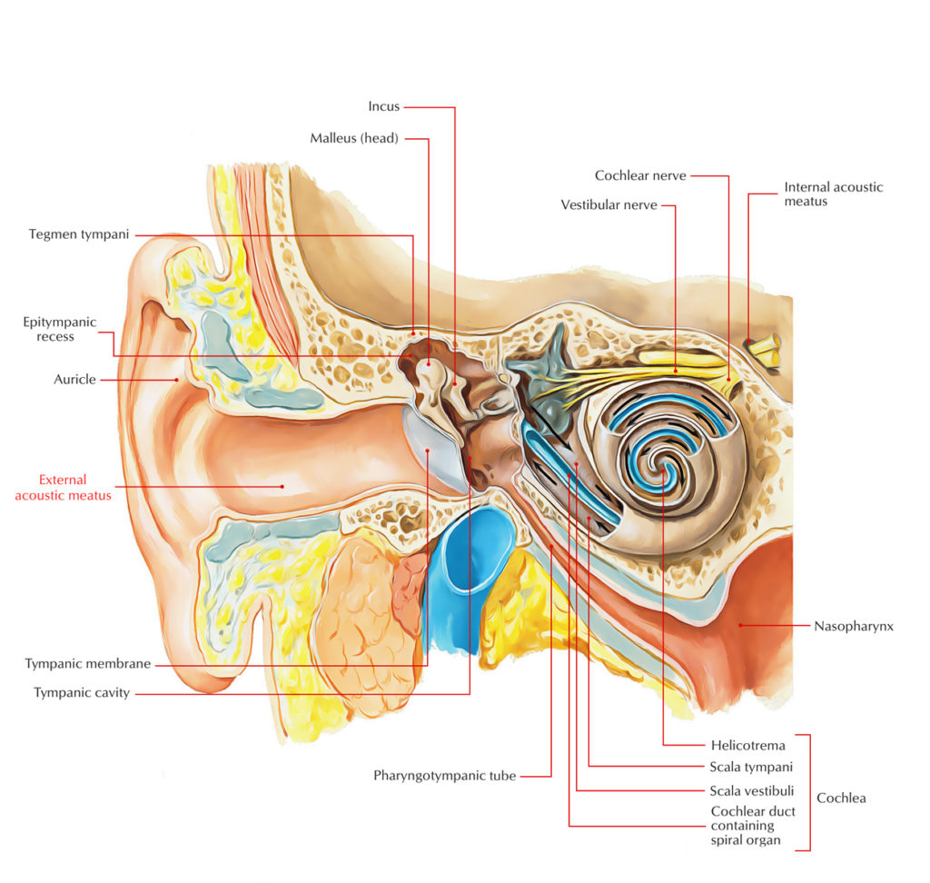
The ear canal ( external acoustic meatus, external auditory meatus, EAM) is a pathway running from the outer ear to the middle ear. The adult human ear canal extends from the pinna to the eardrum and is about 2.5 centimetres (1 in) in length and 0.7 centimetres (0.3 in) in diameter. Structure The human ear canal is divided into two parts.
Hiatus Canalis Nervi Petrosi Majoris Margaret Wiegel™. Mar 2023
The internal auditory canal (IAC), also referred to as the internal acoustic meatus lies in the temporal bone and exists between the inner ear and posterior cranial fossa. It includes the vestibulocochlear nerve (CN VIII), facial nerve (CN VII), the labyrinthine artery, and the vestibular ganglion.
.jpg)
Anatomy Standard Drawing Basis cranii interna dorsolateral view
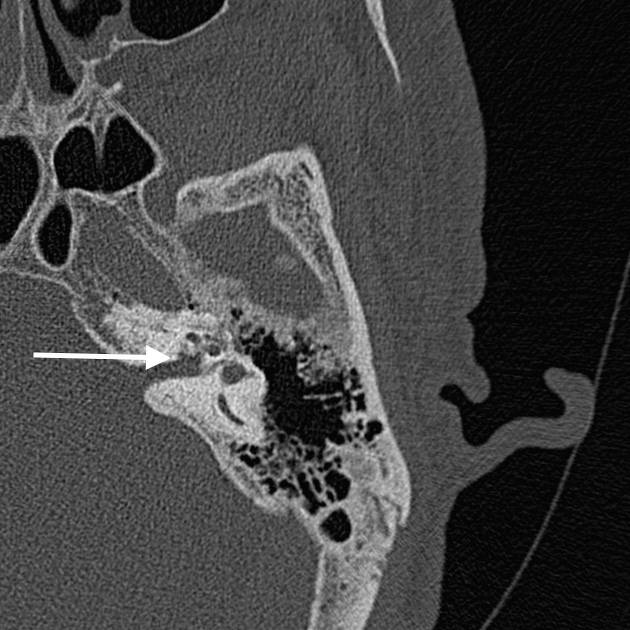
The aim of this paper was to present micro-computed tomography (micro-CT) high resolution images of the fundus of internal acoustic meatus (FIAM) and characterise the normal appearance of its singular areas which are places of passage of numerous anatomical structures. By using micro-CT we obtain detailed volume rendering images presenting topography of the FIAM in 3-dimensional (3D) space.
Divertikel Meatus acusticus internus pacs
Internal ear This mixture of bones, nerves, vessels, membranes, and muscles that make up the ear will be described in this article. Contents External ear Auricle External acoustic meatus Tympanic membrane Muscles of the external ear Vasculature of the external ear Innervation of the external ear Middle ear Tympanic cavity Auditory ossicles

Meatus acusticus internus OUTER EAR
The structure lies in the temporal bone of the cranial cavity and is referred to as the porus acusticus internus. The IAM includes the facial nerve, vestibulocochlear nerve and ganglion, and the labyrinthine artery.. The internal auditory meatus is a critical pathway for important cranial nerves and blood vessels to enter and exit the inner.

Odyoloji Okuyoruz Biz
Özocak O, Unur E, Ülger H, Ekinci N, Aycan K, Acer N. Meatus acusticus internus'un morfometrisi ve varyasyonları. Erciyes Üniversitesi Sağlık Bilimleri Dergisi. 2004;13(3) 1-7. 4. Kobayashi H, Zusho H. Measurements of internal auditory meatus by polytomography. The British Journal of Radiology 1987; 60 (711): 209-14.
Eustachian (pharyngotympanic) tube Anatomy and function Kenhub
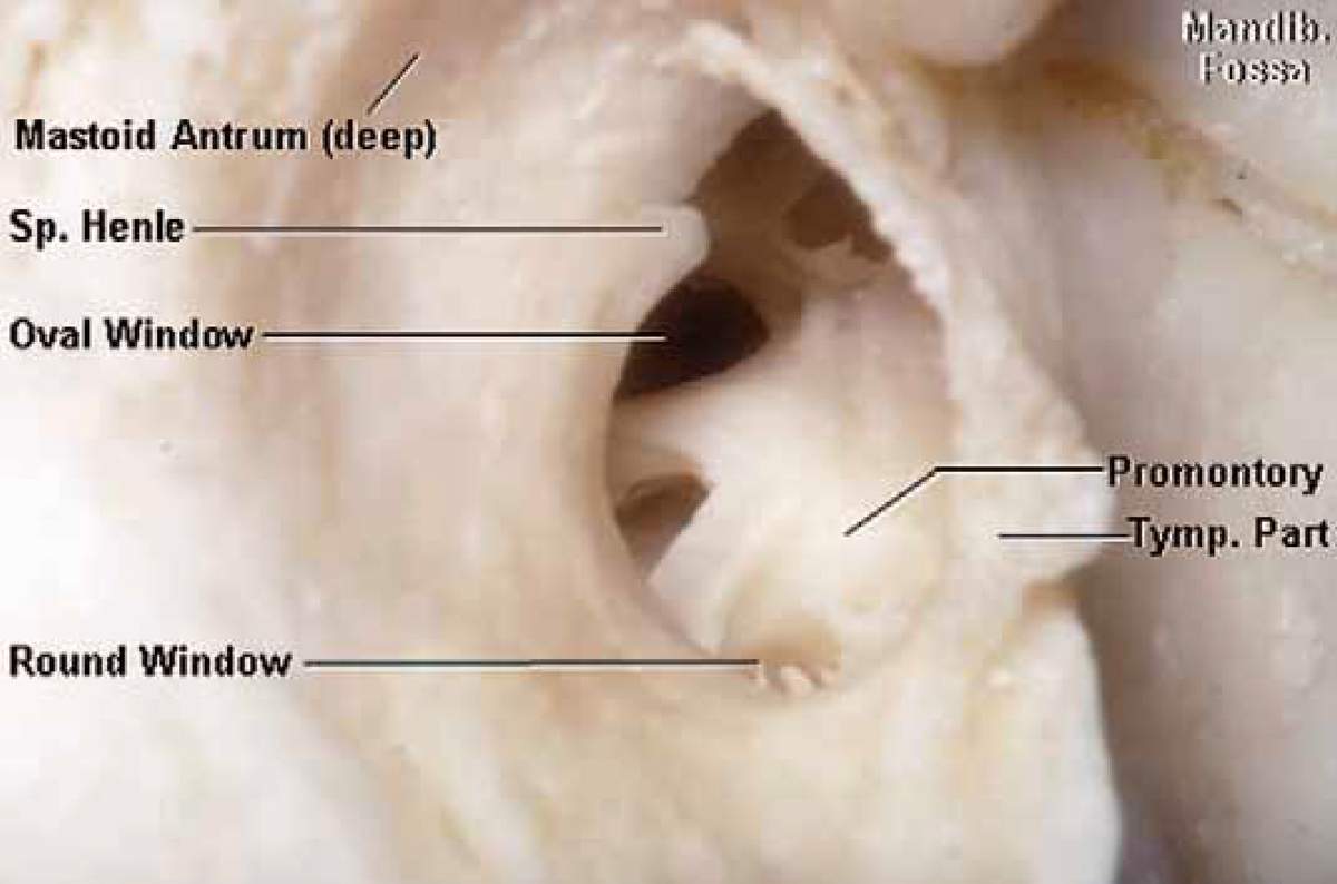
Internal acoustic meatus refers to a small bony foramen situated on the posterior surface of the petrous part of temporal bone, inside the posterior cranial fossa. It allows for the passage of three important structures, namely the vestibulocochlear nerve, facial nerve and the labyrinthine artery.

Meatus acusticus internus WikiSkripta
1. Background. The introduction of computerized transverse axial scanning (tomography) is a milestone in the development of applied radiology (1, 2).Quantitative and morphometric assessment of the internal auditory canal (IAC) are essential to establish the anatomical bases for microsurgery of the cerebellopontine angle and acoustic neuroma, which may produce bone changes and is an important.

PPT ORGANON AUDITIVUS PowerPoint Presentation, free download ID5684974
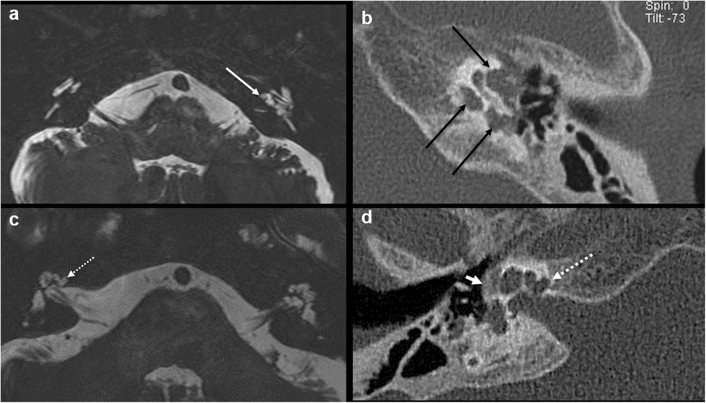
A trumpeted internal acoustic meatus (IAM) is an indirect sign of a vestibular schwannoma and is useful in helping to differentiate between one and other cerebellopontine angle entities, especially from a meningioma which typically does not extend into the meatus and is more often associated with hyperostosis 1.
Innerer Gehörgang Anatomie und Klinik Kenhub
This form of the tumor is located in the internal auditory meatus (meatus acusticus internus), which is a canal within the petrous part of the temporal bone. Besides the vestibular and cochlear nerves (nervus vestibulocochlearis), this canal also channels the facial nerve (nervus facialis) and the gustatory nerve (Chorda tympani).